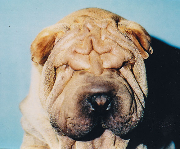
The two most common problems associated with dog eyelids are abnormal conformation and eyelid tumors. Abnormal conformation simply means anything other than a nice, tight-fitting almond-shaped eyelid shape. Many breeds of dogs are bred to have unusual lid conformation, such as bloodhounds, St. Bernards, and flat-faced breeds such as Pugs (see Macropalpebral Fissure Syndrome ). Sometimes the unusual lid conformation leads to medical problems that damage the eye. Some of these conditions are:
1. ENTROPION, which is a rolling-in of the eyelid. This causes the hair on the surface of the eyelid to rub on the eyeball, which is both painful and often causes corneal ulcers or erosions. The corneal damage can also result in corneal scarring, which can interfere with vision. Usually the dog will squint and tear excessively. However, many flat-faced dogs with medial entropion (involving the inside corner of the eyes) show no obvious signs of discomfort.
Entropion is treated by surgical correction (“ blepharoplasty “), which is essentially plastic surgery. Excessive folds and sections of facial skin are removed, and the eyelids tightened. It is uncommon for entropion to recur after surgery unless the entropion is quite involved, particularly in the Shar Pei breed. Very young puppies with entropion will often have “lid tacking” performed (rather than plastic surgery), in which temporary lid sutures are placed to roll out the lids. Often, these puppies do not require permanent plastic surgery once they have matured and “grown into” their facial skin. Permanent plastic surgery is usually not performed in puppies less than 5 or 6 months of age, giving the dog some time to develop its mature head conformation.
Dogs with inherited entropion should not be bred, as they can pass the trait on to their offspring. Veterinary ophthalmologists partner with the Orthopedic Foundation for Animals (OFA) to screen purebred dogs for inherited ocular diseases (see OFA information )
If you suspect that entropion is present in your pet, please consult with your family veterinarian. Your doctor may elect to have your pet referred to a veterinary ophthalmologist for further evaluation and possible surgical treatment.
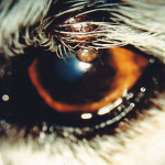
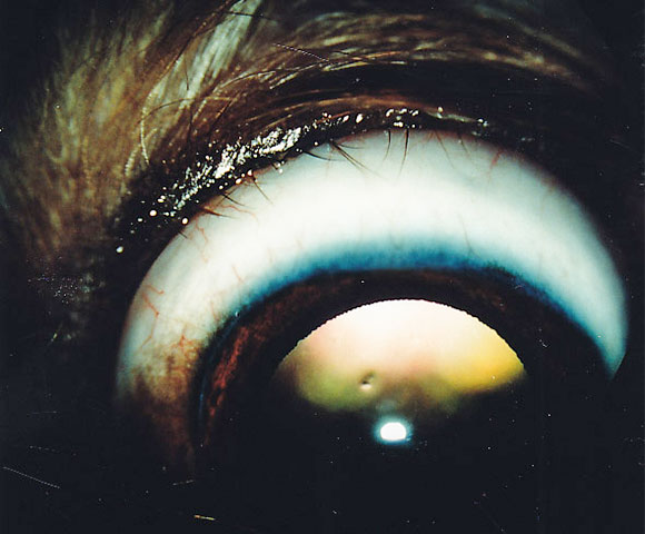
Eyelid tumor in a dog.
2. EYELID TUMORS.
Older dogs commonly develop eyelid tumors (cancer). As in humans, cancer can be either benign or malignant. Fortunately, eyelid tumors in dogs are usually benign and do not spread to distant tissues. However, eyelid tumors do slowly or quickly grow, and can destroy the structure of the eyelid, in addition to rubbing on the eye. It is usually best to remove them when they are still small.
Eyelid tumors are treated by surgical removal. While there are many different surgical procedures possible, most eyelid tumors in old dogs can be removed at Animal Eye Care without requiring general anesthesia. The patient is given a sedative, and then a local eyelid anesthetic is given to numb the eyelid. The tumor is removed and the site frozen with liquid nitrogen (cryosurgery) to kill any remaining tumor cells. Tumor cells are usually very sensitive to freezing, and normal eyelid tissue is more resistant. After surgery, the eyelid margin turns pink (depigments), but usually repigments within 4 months.
It is rare for the eyelid tumor to recur following surgery. 85-90% of tumors do not recur following surgery.
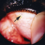
The arrow points to a site of ectopic cilia on the inside surface of the upper lid of this young Golden Retriever.
3. DISTICHIASIS AND ECTOPIC CILIA.
Eyelids of dogs can grow abnormal hairs. These hairs grow from the oil glands (Meibomian glands) of the lids and are called distichia if the hair protrudes from the oil gland opening onto the edge of the eyelid. Distichia are often irritating, especially if the hairs are long and stiff. Ectopic cilia are also hairs growing from oil glands on the eyelid, but the hair protrudes from the inner surface of the eyelid and is very painful, often causing corneal ulcers.
Dogs with distichiasis may or may not show signs of discomfort, ranging from slight intermittent squinting and/or rubbing of the eyes, to severe squinting and discomfort. Dogs with ectopic cilia are always uncomfortable. Most dogs with ectopic cilia are young adult dogs or older puppies. Both conditions are common in Shih Tzus. Many other breeds have problems with distichia. At Animal Eye Care, both conditions are treated surgically under general anesthesia, with a procedure called cryoepilation. With this procedure, the abnormal hair follicles are frozen using a liquid nitrogen probe, and the hairs then removed.
Numerous distichia rubbing on the surface of the eye.
After surgery, the eyelids are swollen for 4-5 days, and the eyelid margins will depigment and turn pink. Usually, the lid margins will repigment within 4 months. It is important to understand that new abnormal hairs can grow from new sites after surgery, but this is uncommon in dogs older than 3 years old (unless the dog is a Shih Tzu). With cryoepilation, most (if not all) of the treated hair follicles do not regrow (unless the dog is a Shih Tzu). Repeat surgical treatment is rarely required, unless the animal is a puppy (and grows new hairs in new sites) or a Shih Tzu.
4. PROLAPSED THIRD EYELID GLAND (PTEG) . This condition is also called Prolapse of the Nictitans Gland. A slang term (which is to be discouraged) is “cherry eye”. Dogs have a third eyelid that slides up over the surface of the eye for protection. The third eyelid also has a tear gland located deep within its tissues, called the third eyelid gland TEG). Each eye of a dog actually has 2 tear glands (also called lacrimal glands), unlike people (who have one). The orbital lacrimal gland produces 60% of the tears for the eye, and the third eyelid gland produces 40% of the tears. The TEG has a T-shaped piece of cartilage in it, and is hidden out of sight and anchored to the tissues of the eye socket by ligaments. Some dogs are born with weak ligaments, which allow the TEG to pop out of its normal position and look like a pink roundish object in the inside corner of the eye.
PTEG is suspected to be inherited in some breeds, as it occurs with increased frequency in some breeds, notably the American Cocker Spaniel, Lhasa Apso, and English Bulldog. The condition can be in one eye or both eyes, and if in both eyes, it can occur weeks to months apart. Treatment is surgical, and involves repositioning the PTEG and suturing it into place. The prolapsed TEG should not be removed! If the condition is left untreated, the eye is at great risk for developing dry eye months to years later. Additionally the PTEG can swell and be uncomfortable, and interfere with vision. However, while surgery decreases the chances that the eye will develop a dry eye problem, it does not eliminate this risk.
The success rate of surgery is approximately 95% for non-recurrence of the PTEG, except in the English Bulldog and Mastiff breeds, in which the success rate is lower.
If you suspect that your pet has PTEG, please consult with your family veterinarian. Your doctor may recommend referral to a veterinary ophthalmologist for evaluation and surgical treatment.