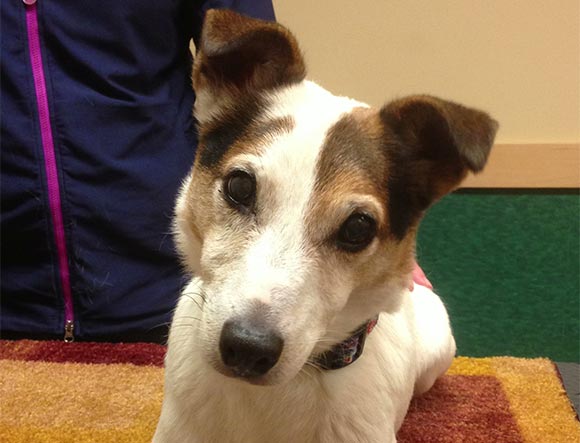
9 years ago, this dog’s eye was terribly injured by a cat claw when the dog was a tiny puppy. The claw perforated the cornea, ruptured the lens, and caused a retinal detachment. The eye was blind but VERY luckily did not need to be removed. Thanks to lifetime regular ophthalmic care, it has remained a cosmetic, blind eye. Can you tell which eye was injured?
Ocular injury is unfortunately a common occurrence in animals. Many kinds of injuries occur, such as eyelid lacerations, proptosis (where the eye is forced out of the eye socket), blunt injury to the eye and/or eyelids, thorn or other foreign body injuries, cat claw injuries, dog bite injuries, lacerations of the cornea, gunshot pellet wounds, and severe head/eye socket injury from being kicked by a horse, or hit by a car. Other injuries can be chemically induced, such as mace sprayed into the eyes, or paint or soap burns of the cornea. Still other injuries can occur secondary to periocular insect or spider bites.
The key to any eye injury is to seek immediate veterinary attention and prevent further trauma to the eye. The injured pet often wants to rub the eye, and this must not be allowed. It is a good idea to have a cone shaped restraint collar (E collar) on hand, that has been custom fitted for your pet. You can purchase this from your family veterinarian, and it can also be used in other emergencies, to prevent your pet from traumatizing other parts of its body, such as the hindquarters. It is also a good idea to have a bottle of eye irrigating solution on hand, such as sterile saline solution (NOT contact lens cleaning solution) to irrigate the eye, especially if the eye is proptosed (pushed out of the socket). Seeking immediate veterinary assistance and placing an E collar on the pet can make the difference between a blind eye and a visual eye, or whether or not the eye needs to be removed. Often, however, the injury is so severe that no matter how soon the pet is treated by a veterinarian, the eye cannot be saved. It is a good idea to ask your veterinarian if they think your pet should be referred to a veterinary ophthalmologist for evaluation, if you are willing to be referred to a specialist.
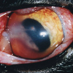
Ruptured eye of cat, from cat claw injury.
One of the most frequent types of eye injuries encountered is “New Puppy Meets Cat.” It only takes a fraction of a second for a cat to decide whether or not the puppy is a friend or an adversary, and in this time span, the paw lashes out towards the bright, shiny puppy eye. A cat claw injury is often quite serious, but initially may not seem to be. Cat claws are filthy, and when a claw injures an eye it also delivers a large load of bacteria to the wound. Secondary infection can develop, and if the lens were ruptured by the claw, the lens rupture might lead to surgical removal of the eye, unless the injured lens is removed as soon as possible by a veterinary ophthalmologist. Even if the lens were removed, the eye is still at high risk for nonreturn of vision. If the cat claw lacerated the cornea alone, suturing the wound closed by a veterinary ophthalmologist is often successful in saving the eye.
Another frequent eye injury is from dog bites . Often, a puppy is on the receiving end, having strayed too close to the bigger dog’s food bowl. Often, the eye is proptosed or at the very least; very bruised and contused. If the eye is “squeezed” by the dog bite, it may look relatively OK immediately after the injury, but in reality the eye is severely damaged internally, and retinal detachment, hemorrhage, and/or glaucoma may occur, not to mention cataract and/or lens rupture. Think of the eye as a soft ball. If the ball were punched hard, it would deeply indent quickly, and then quickly return to its round shape. Eyes are very sensitive, and if injured in this manner, irreparable damage can occur in a hurry.
Proptosis is another frequent eye injury in dogs, particularly flat-faced dogs. Pugs, Shih Tzus, and Pekingese all have flat faces and shallow eye sockets. The shallow sockets and the large eyelid openings make them high risk for proptosis occurring (see Macropalpebral Fissure Syndrome ). An eye forced from the eye socket is an emergency, and must be replaced in the eye socket by a veterinarian as soon as possible. Until you can reach a veterinarian, keep the eye moist with sterile saline solution (NOT cleaning solution), eye irrigating solution, or (as a last resort) water. If you have any artificial tears, this also can be helpful, but it is important to keep the tissues moist and the patient quiet. Do not give aspirin or any other pain medication — let the doctor decide what type of analgesic to give. Do not let the dog rub at its eye, and take away its food and water, as the dog will likely be anesthetized in order to have the eye replaced in the eye socket. Even after the eye is replaced, there is a significant risk that the eye will not recover vision, and might eventually require surgical removal (enucleation). Sometimes the proptosed eye can be saved, but needs plastic surgery to protect it (medial canthoplasty).
If the eye trauma is so severe that the eye must be removed (enucleation) , there are two methods to remove the eye. One is a standard enucleation, in which the eye is removed and the eyelids permanently sutured closed. The animal does not miss this eye psychologically, unlike the owner. This surgery can be performed by general practitioner veterinarians, and does not usually require a veterinary ophthalmologist to perform the surgery. However, there is a second method to remove the eye, which is more cosmetic, called Enucleation and Placement of an Orbital Prosthesis. This surgery is performed by a veterinary ophthalmologist. The eye is removed, but just before the lids are permanently sutured closed, a sterile prosthetic ball is placed in the eye socket, and then the lids are sutured closed. This hidden ball (orbital prosthesis) prevents the lid skin from sinking down into the eye socket (which would be the case with a standard enucleation), and makes the pet look like the eye is simply closed. Animals in which the orbital prosthesis is not recommended: patients with an infected eye socket, and patients with extremely flat faces and very shallow eye sockets. With both surgical methods, healing time is usually two weeks, and it is best if a cone shaped restraint collar (E collar) is placed on the animal postoperatively to prevent self-trauma.
This informational article cannot possibly address all the different types of eye trauma in domestic animals. Keep in mind that each injury is unique, and treatment for that injury is specific for that particular eye and patient.